Cerebral Aneurysm
- A cerebral aneurysm (brain aneurysm) is a weak spot of a blood vessel (artery) in the brain that bulges out.
- Saccular aneurysm, also referred to as a “berry aneurysm” is a round sac that comes off the artery.
- Fusiform aneurysm, is an aneurysm that bulges on all sides of the artery.
- Brain aneurysms are frequently diagnosed incidentally during workup for other medical conditions.
- Risk factors for developing a brain aneurysm include genetics such as connective tissue disorders that weaken the blood vessels or family history, polycystic kidney disease, and cigarette smoking.
- Risk factors related to aneurysm rupture include untreated high blood pressure, smoking, aneurysm size, and family history.
- Brain aneurysms can become symptomatic if they are large, compressing a nerve, or prior to rupture. Symptoms include headaches, double vision, extremity weakness or sensory changes.
- Possible treatments for cerebral aneurysms include coiling, stenting, flow diversion, or clipping. Some aneurysms are eligible for close monitoring without treatment.
- If a brain aneurysm ruptures patients develop a special pattern of bleeding in the brain, also known as subarachnoid hemorrhage. In that setting patients can present with sudden “worst headache of life”, vision changes, sensitivity to light, nausea/vomiting, loss of consciousness, or weakness in extremities.
- If you have any of these symptoms, call 911 immediately to seek emergent medical care.
Stroke
- An ischemic stroke happens when an artery in the brain is blocked by a blood clot causing diminished blood flow and oxygen to that area of the brain.
- An ischemic stroke can occur when a plaque ruptures from your carotid arteries, or when a blood clot travels from elsewhere in the body, usually the heart.
- Modifiable risk factors include high blood pressure, diabetes, high cholesterol, cigarette smoking, sedentary lifestyle, and obesity.
- Non-modifiable risk factors include age, race, sex, and genetics.
- Stroke like symptoms that resolve within 24 hours, without residual deficits, constitute a transient ischemic attack (TIA).
- Treatment of ischemic stroke includes clot busting medications (IV-tPA) and mechanical thrombectomy.
- A hemorrhagic stroke occurs when a blood vessel bursts causing bleeding in the brain.
- A hemorrhagic stroke is caused by high blood pressure, AVM or other vascular malformation, cerebral aneurysm, or the use of blood thinning medications.
- Symptoms of an ischemic or hemorrhagic stroke include but not limited to acute onset of trouble speaking, facial droop, weakness or numbness in extremities, dizziness, double vision, or difficulty walking.
- If you have any of these symptoms, call 911 immediately to seek emergent medical care.
Arteriovenous Malformation (AVM)
- Arteriovenous malformation (AVM) is an abnormal tangle of blood vessels that has an irregular connection between the arteries and veins in the brain or spine. The arteries (high pressure system) are directly connected to the veins (low pressure system) without the middle connection, the capillary system, which normally decreases the pressure of the blood.
- Possible treatments for AVMs include onyx embolization, surgical resection, or stereotactic radiosurgery. Some AVMs are eligible for close monitoring without treatment.
- Over time the blood vessels within the AVM become weakened causing rupture and bleeding in the brain.
- If an AVM or arteriovenous fistula is located in the spine it can cause progressive worsening symptoms over several months, which can be mistakenly attributed to degenerative spine disease.
- If an AVM ruptures, symptoms include headache, seizures, weakness in extremities, vision changes, dizziness or speech difficulties. However, symptoms are variable depending on the location of the AVM.
- If you have any of these symptoms, call 911 immediately to seek emergent medical care.
Cerebral Cavernous Malformation (CCM)
- Cerebral Cavernous Malformation or Cavernoma (CCM) is a collection of small blood vessels (capillaries) in the brain that are enlarged and irregular in structure. These capillaries have abnormally thin walls that are prone to leak. They also lack other support tissues , such as elastic fibers, which normally make them stretchy. As a result, when the capillaries fill with blood, they stretch out and create "caverns." They may not return to their normal size when the blood vessels empty.
- Most CCM are the result of a genetic mutation that sometimes can be passed on from one generation to the next. Some patients present with multiple CCMs.
- Possible treatments for CCM include surgical resection, or stereotactic radiosurgery. Some CCMs are eligible for close monitoring without treatment.
- Over time these blood vessels can rupture and cause bleeding in the brain.
- If a CCM is located in the spine it can cause progressive worsening symptoms over several months, which can be mistakenly attributed to degenerative spine disease.
- If a CCM ruptures, symptoms include headache, seizures, weakness in extremities, vision changes, dizziness or speech difficulties. However, symptoms are variable depending on the location of the CCM.
- If you have any of these symptoms, call 911 immediately to seek emergent medical care.
Carotid Artery Stenosis
- Carotid artery stenosis is narrowing of the vessel due to atherosclerotic plaque caused by fat, cholesterol, and calcium. The carotid artery is located on each side of the neck and perfuses the front part of your brain.
- Carotid artery stenosis is usually diagnosed by ultrasound, CTA (computed tomography angiography), MRA (magnetic resonance angiogram) or cerebral angiogram.
- Risk factors include high blood pressure, diabetes, high cholesterol, cigarette smoking, sedentary lifestyle, and obesity
- When pieces of the plaque break off, clots are formed, which can block blood vessels in the brain. This results in a stroke or a transient ischemic attack.
- Possible treatments for carotid artery stenosis include carotid endarterectomy and carotid artery stenting. Some patients with carotid disease are eligible for close monitoring (typically with ultrasound or other non-invasive imaging modality) and medical management through risk factor control.
- If the carotid artery stenosis becomes severe, patients are at higher risk of stroke or transient ischemic attack. Symptoms include trouble speaking, facial droop, weakness or numbness in extremities.
- If you have any of these symptoms, call 911 immediately to seek emergent medical care.
Intracranial Atherosclerotic Disease (ICAD)
- Intracranial atherosclerotic disease (ICAD) is narrowing of the vessel due to atherosclerotic plaque caused by fat, cholesterol, and calcium. These arteries provide blood to various parts of your brain.
- ICAD is usually diagnosed by transcranial ultrasound, CTA (computed tomography angiography), MRA (magnetic resonance angiogram) or cerebral angiogram.
- Risk factors include high blood pressure, diabetes, high cholesterol, cigarette smoking, sedentary lifestyle, and obesity
- When pieces of the plaque break off, clots are formed, which can block blood vessels in the brain. This results in a stroke or a transient ischemic attack.
- Possible treatments for ICAD include medications (usually in the form of aspirin or plavix) and intracranial angioplasty and stenting. Most patients with ICAD are eligible for close monitoring (typically with ultrasound or other non-invasive imaging modality) and medical management through risk factor control.
- If ICAD becomes severe or causes symptoms, patients are at higher risk of stroke or transient ischemic attack. Symptoms include trouble speaking, facial droop, weakness or numbness in extremities.
- If you have any of these symptoms, call 911 immediately to seek emergent medical care.
Moyamoya Disease
- Moyamoya disease is a cerebrovascular disorder that affects the blood vessels in the brain causing them to narrow or constrict limiting the flow of oxygen and blood. Other blood vessels are formed to increase circulation to the affected area, however these vessels are small and become easily occluded.
- Moyamoya means “puff of smoke” in Japanese and describes the look of the blood vessels on angiographic imaging.
- The cause of the disease is unknown however is more common in Japan, Korea, and China.
- Risk factors include familial history, Asian decent and female.
- Moyamoya is diagnosed during workup for cause of a stroke or recurrent transient ischemic attacks.
- Possible treatment for Moyamoya disease includes EC-IC bypass. Some patients with Moyamoya disease are eligible for close monitoring without treatment.
- Symptoms are those of a stroke including but not limited to trouble speaking, facial droop, weakness or numbness in extremities, dizziness, or difficulty walking.
- If you have any of these symptoms, call 911 immediately to seek emergent medical care.
Trigeminal Neuralgia
- Trigeminal neuralgia is characterized by chronic debilitating facial pain. The pain can involve the eye, cheek, jaw, or the entire face. Your face becomes extremely sensitive to the touch and the pain is similar to an electric shock.
- One of the most common causes of trigeminal neuralgia is the compression of the nerve that controls sensation to the face (trigeminal nerve) by an artery in the brain.
- Trigeminal neuralgia is diagnosed based on clinical symptoms, although an MRI is usually obtained to identify the responsible blood vessel.
- Possible treatments for trigeminal neuralgia include medications, percutaneous procedures, stereotactic radiosurgery, or microvascular decompression.
Hemifacial Spasm
- Hemifacial spasm is characterized by chronic debilitating involuntary twitching of the face. Twitching can involve the eye, cheek, jaw, or the entire face. It can often be difficult to keep your eye open or to read a book.
- One of the most common causes of hemifacial spasm is the compression of the nerve that controls movement of the face (facial nerve) by an artery in the brain.
- Hemifacial spasm is diagnosed based on clinical symptoms, although an MRI is usually obtained to identify the responsible blood vessel.
- Possible treatments for hemifacial spasm include medications, Botox injections, or microvascular decompression.
Intractable Epistaxis
- Intractable epistaxis is characterized by severe uncontrolled nose bleeds that can sometimes become life threatening.
- Frequent causes include facial trauma, foreign bodies, nasal or sinus infections, and prolonged inhalation of dry air.
- Risk factors include use of blood thinners, congestive heart failure, diabetes mellitus, hypertension, and a history of anemia
- In most patients with epistaxis, the bleeding responds to cauterization, nasal packing, or both. In refractory cases, some patients might benefit from endovascular particle embolization.
- If you develop a severe nose bleed, call 911 immediately to seek emergent medical care.
Idiopathic Intracranial Hypertension (IIH)
- Idiopathic Intracranial Hypertension (IIH) or pseudotumor cerebri is the result of elevated pressure inside the skull, due to built up of cerebrospinal fluid (CSF). This develops spontaneously and is not the result of other diseases such as brain tumors. Symptoms include persistent headaches and blurry vision.
- IIH is usually diagnosed by a lumbar puncture that demonstrates high pressure.
- Most patients with IIH have stenosis in one of the venous sinuses of the brain, which results in limited drainage of CSF and build up of pressure.
- Possible treatments for IIH include medications, ventriculoperitoneal shunt, and venous sinus stenting. Most patients with IIH are eligible for close monitoring (typically with frequent eye exams) and medical management.
- If IIH becomes severe, patients are at higher risk of vision loss.
- If vision loss develops, call 911 immediately to seek emergent medical care.
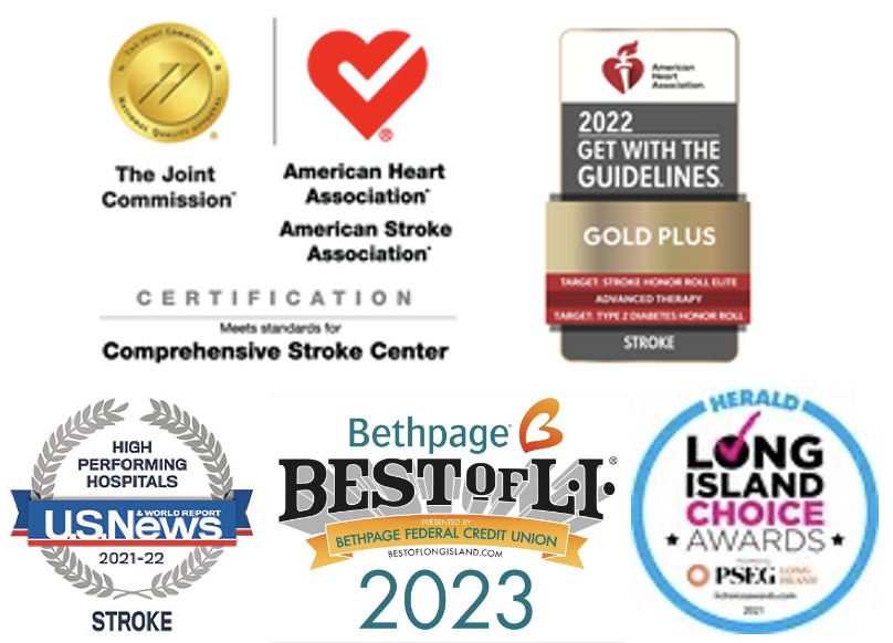
abOUT USThank you for your interest in The Stroke & Brain Aneurysm Center of Long Island®. Our program has attracted a diverse group of national experts, and is the first and only Joint Commission Certified Comprehensive Stroke Center in the South Shore of Long Island.
|
outpatient locationsBabylon
60 George Street Babylon, NY 11702 West Islip 380 Montauk Highway West Islip, NY 11795 Smithtown St. Catherine of Siena Medical Office Bldg. 48 Route 25A, Suite 302 Smithtown, NY 11787 |
inpatient LOCATIONTel: 631-983-7072
Good Samaritan University Hospital 1000 Montauk Hwy. West Islip, NY 11795 http://goodsamaritan.chsli.org/strokebraincenter |
The Stroke and Brain Aneurysm Center of Long Island © All rights reserved 2024